Anatomy of the Drosophila adult midgut
1- Anatomical description of the digestive tract
The
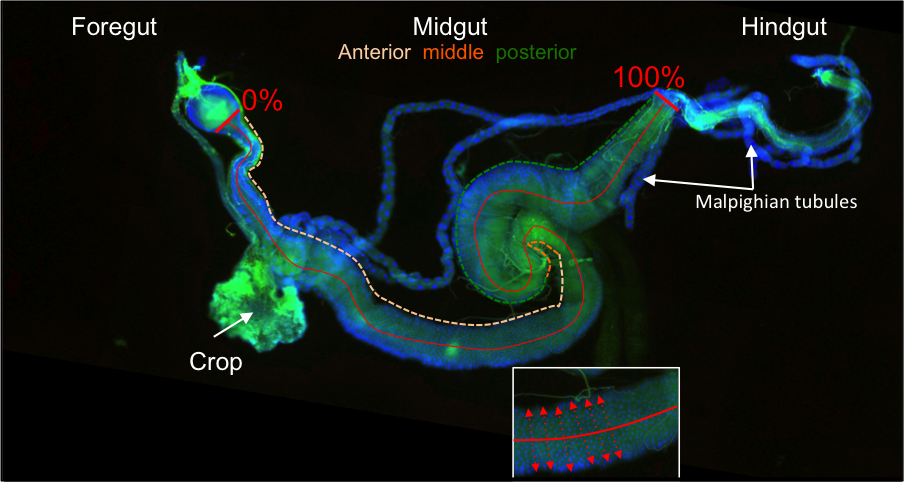
Drosophila adult gut is composed of three main parts: a- the foregut, comprising the oral cavity, the esophagus, the Crop (depicted in the photo below), and the cardia; b- the midgut; and c- the hindgut. The fore- and hindgut have an ectodermal origin, whereas the midgut has an endodermal origin. The cardia is a valve-like organ at the junction between the foregut and the midgut that secretes the peritrophic matrix. The cardia is separated in two parts: the ectodermal proventriculus and an endodermal part that constitutes the most anterior part of the midgut (that we named R0). Malpighian tubules, which play a role analogous to the vertebrate kidneys, connect the gut at the midgut-hindgut junction.
To determine the position of each region, we normalized the midgut length to 100% starting around the middle of the cardia (where the gut tube is composed only of endoderm: see photo) and ending at the midgut-hindgut junction, where Malpighian tubules branch.
2- Morphological analysis of the Drosophila midgut
a- Morphometric analysis of the midgut
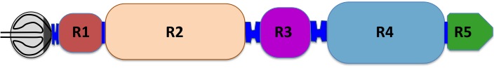
We measured the radius of the gut along its length. Five major constrictions (hereafter referred to as anatomical boundaries) were found to separate the midgut into chambers that we named Regions 1-5 (R0 corresponds to the endodermal part of the cardia that we excluded from our analysis).
|
b- Organization of the midgut in 3D
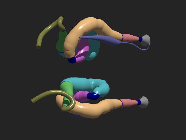
Based on gut morphometric measurements and histological sections of whole flies, we reconstructed a 3D model of the midgut. The set of points defining the gut model can be found
here. Then, different types of features can be mapped onto this surface and rendered in user-frienly 3D views (such as anatomical features, see below;
histological features or
gene expression pattern features). We then projected the five regions and their boundaries on the 3D reconstruction. The 3D view reveals that boundaries are located to specific anatomical coordinates: BR1-R2 is at the junction thorax abdomen, BR2-R3 is the most ventral point of the gut, BR3-R4 is the most dorsal point of the gut, and BR4-R5 marks the beginning of the midgut ascension towards the hindgut. Altogether, our anatomical data indicate that:
- R1 is a region located in the thorax and ends at a position that corresponds to an anatomical hub where the midgut, the crop, and malpighian tubules physically interact.
- R2 corresponds to an abdominal part of the midgut that is dorsally located and that falls back ventrally at the posterior part. R1 and R2 correspond to the previously described "anterior midgut".
- R3 corresponds to the previously described region called "middle midgut" and contains the copper cells. This region initiates a retrograde folding of the midgut, (from posterior to anterior), and it follows a ventral-dorsal axis.
- R4 forms a complex loop that re-orients the gut towards the posterior part of the abdomen and ends ventrally.
- R5 corresponds to an ascending segment of the gut that joins the hindgut. R4 and R5 form the previously characterized posterior midgut.
Click on the picture below to activate the 3D view.
List of anatomical structures
| Color |
Name |
Position |
Histological regions |
Sub-regions |
|
Crop |
1 |
Crop |
Crop |
|
Cardia |
2 |
Cardia |
cardia (midgut part), cardia (proventriculus) |
|
Boundary P-1 |
3 |
Boundary P-1 |
Boundary P-1 |
|
R1 |
4 |
R1a, R1b |
R1a, R1b |
|
Boundary 1-2 |
5 |
NA |
NA |
|
R2 |
6 |
R2 |
R2a, R2b, R2c |
|
Boundary 2-3 |
7 |
Boundary 2-3 |
Boundary 2-3 |
|
R3 |
8 |
R3ab, R3c |
R3a, R3b, R3c |
|
Boundary 3-4 |
9 |
Boundary 3-4 |
Boundary 3-4 |
|
R4 |
10 |
R4a, R4bc |
R4a, R4b, R4c |
|
Boundary 4-5 |
11 |
NA |
NA |
|
R5 |
12 |
R5 |
R5a, R5b |
|
Boundary 5-H |
13 |
NA |
NA |
|
Hindgut |
14 |
Hindgut |
Hindgut |
|
|
3D model of regions
|
|
|
|
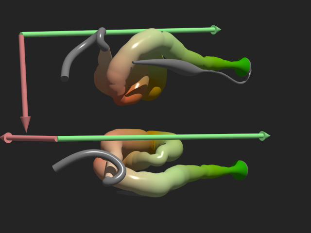
3D model with morphology axes
|
|
|
The dorso-ventral axis and the antero-posterior axes are shown in the following 3D representation:
|
