
| Home | Overview of gut regions | Anatomy | Histology | Transgene expression mapping | Gene expression |
Histology of the Drosophila adult midgut
In this section, we describe the histological diversity along the Drosophila midgut. This atlas is based on transversal sections of the different gut regions that were marked with either GFP (immunostaining) or classical histological stains. Below is shown:- A summary showing the different histological territories of the midgut in 3D with links to representative sections of each region (click on "Show" on the right).
- A schematic representation of the histology of the midgut regions and representative sections of each region.
- A series of 34 histological sections throughout the midgut.
- Two videos showing the overall organization of the Drosophila adult midgut.
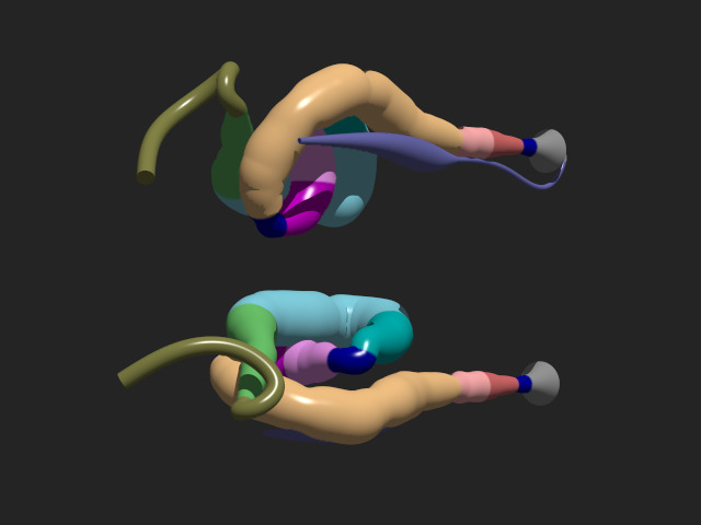
1- Histological territories in 3D and representative sections of each region.
Click on "Show" on the right of the table to see representative sections of each region. To activate the 3D view, click on the picture.
|
|
2- A schematic representation of the histology of the midgut regions and representative sections of each region.
By clicking on the region of interest, you will find a scheme that summarizes the histological data of the region, as well as representative images of histological gut sections. Methods to stain the gut can be found in the Supplementary Material and Methods section of our manuscript. We used in this analysis:- a marker of brush borders (GFP transgene A142)
- a marker of actin revealing visceral muscles (phalloidin)
- a marker of mucopolysaccharides and glycosaminoglycans (alcian blue)
- a marker of cell junctions (E-cad)
- a marker of the apico-lateral compartment (dlg)
- a marker of nuclei (DAPI)
- a marker of lipid particles (oil redo staining)
- a marker of polysaccharides (Periodic acid Schiff or PAS)
- a counter-staining with hematoxylin
blabla
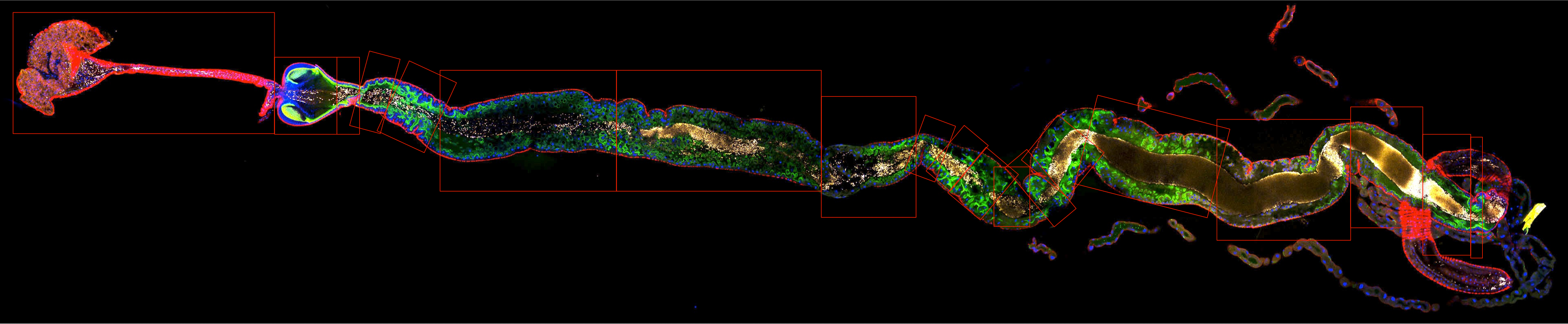
3- 34 histological sections throughout the midgut
This slideshow presents successive histological sections along the gut with their longitudinal positions.blue: nuclei stained with DAPI; green: brush borders stained with the A142 GFP trap, red: visceral muscles stained with phalloidin; yellow: food particles.
Link to slideshow
4- Two videos showing the overall organization of the Drosophila midgut.
|
3 color video of the gut showing its internal and external organization: The midgut of a 3-5 day-old adult female was dissected and imaged by confocal microscopy in xyz axis, and then reconstructed in 3D using Imaris. blue: nuclei stained with DAPI; green: brush border marked with the A142 GFP trap; red: food. | Download high-resolution movie |
|
4 color video of the 3D gut structure: The midgut of a 3-5 day-old adult female was dissected and imaged by confocal microscopy in xyz, and then reconstructed in 3D using Imaris. blue: nuclei stained with DAPI; green: brush border marked with the A142 GFP trap; red: visceral muscles stained with phalloidin; yellow: food particles (fluorescent latex beads). | Download high-resolution movie |